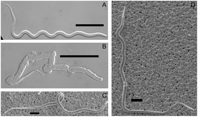
Figure 1. Enzyme sensitivity and external morphology of midgut-derived B. pahangi mf.
Panel A, LVP-derived mf with sheath removed by papain treatment; B, Cpp-derived mf after papain treatment; C, scanning electron micrograph of sheathed LVP-derived mf; D, scanning electron micrograph of sheathed Cpp-derived mf. Scale bars: panels A and B, 50 µM; C and D, 20 µM.

