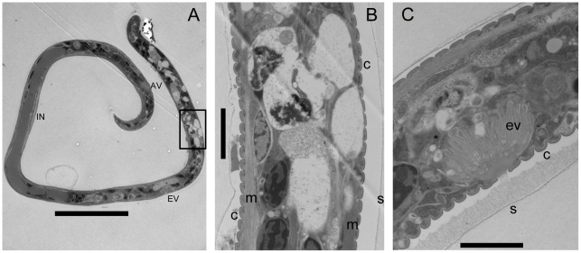
Figure 3. Ultrastructural aspects of Cpp-derived B. pahangi mf.
Longitudinal section demonstrates vacuolization of the nuclear column, disruption of the hypodermis and body wall muscle, and release of material from the excretory vesicle. Panel A, longitudinal section of full length mf; B, high magnification view of nuclear column in the boxed area anterior to the excretory vesicle; C, excretory vesicle activity from a Cpp-damaged worm, showing release of visible material from the pore and accumulation of the material between the scalloped cuticle and the overlying sheath. NR, nerve ring; EV, excretory vesicle; IN, innenkorper; AV, anal vesicle; C, cuticle; m; longitudinal muscle. Scale bars: panel A, 20 µM; B and C, 2 µM.

