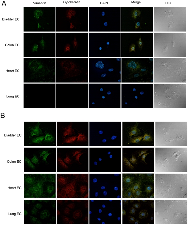
Figure 5. Vimentin and cytokeratin are present on the cell surface.
Different organ derived endothelial cells were incubated with anti-pan-CK (red) or anti-vimentin (green) specific antibodies. In (a), antibodies were incubated with live cells and in (b), the antibodies were incubated with p-formaldehyde fixed and detergent permeabilized cells. DAPI staining (blue), merged fluorescence images and the corresponding DIC images are also shown.

