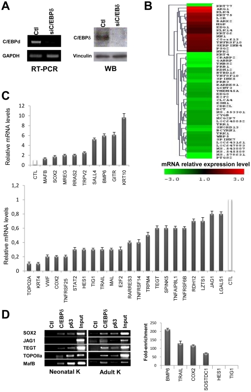
Figure 3. Identification of C/EBPδ-regulated genes in human keratinocytes.
A. Left Panels, RT-PCR analysis of human primary keratinocytes after C/EBPδ RNAi inactivation at 48 hours post-transfection. cDNA normalization was performed with GAPDH. Right Panels, Western Blot analysis of the same C/EBPδ-inactivated human primary keratinocytes with the C/EBPδ antibody. Vinculin was used as a loading control. B. Heat map showing the mRNA expression levels of several classes of genes after C/EBPδ inactivation. C. qRT-PCR analysis of C/EBPδ regulated genes that emerged from the expression profiling. D. ChIP analysis of promoter regions of C/EBPδ-regulated genes with chromatin from human primary keratinocytes, with α-C/EBPδ and control antibodies. In the Left Panel, adult and neonatal keratinocytes were analyzed in semi-quantitative PCR. In the Right Panel, the targets were validated by qPCR in adult keratinocytes.

