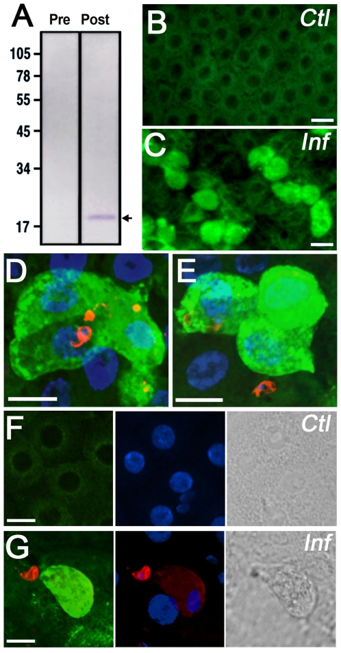
Figure 4. AgApoLp-III protein detection and subcellular localization in control and Plasmodium-infected midguts.
(A) Western blot analysis of hemolymph from adult females probed with pre-immune or polyclonal anti-AgApoLp-III antiserum. A single band of expected molecular weight (19 kDa) for the mature protein was detected and is indicated by the arrow. (B–G) Subcellular localization of AgApoLp-III (green) in control (B and F) and infected (C, D, E, and G) midguts. The ookinete surface was stained using anti-Pb28 monoclonal antibodies (red) and nuclei with DAPI (blue). Phase contrast images of the midgut surface in control (F) and infected (G) midguts are shown in the right panels.

