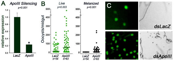
Figure 5. Effect of AgApoLp-III silencing on Plasmodium berghei infection in Anopheles gambiae (G3 strain) females.
(A) Relative abundance of AgApoLp-III mRNA in G3 mosquitoes injected with AgApoLp-III dsRNA (ApoIII) or control LacZ dsRNA (LacZ). (B) Effect of AgApoLp-III silencing on the number of live oocysts (green dots;  ) and melanized parasites (black dots;
  ) in midguts analyzed seven days post infection. Dots represent the number of parasites present on individual midguts, and the median number of parasites is indicated by the horizontal line. Distributions are compared using the Kolmogorov-Smirnov test; n = number of mosquitoes, * indicates significant decrease in mRNA levels relative to dsLacZ-injected controls. (C) Representative field to illustrate the effect of AgApoLp-III silencing on Plasmodium infection 7 days post-feeding. Live oocysts express GFP and exhibit green fluorescence (right panels), while melanized parasites can be observed as dark spots in bright field images (left panels).

