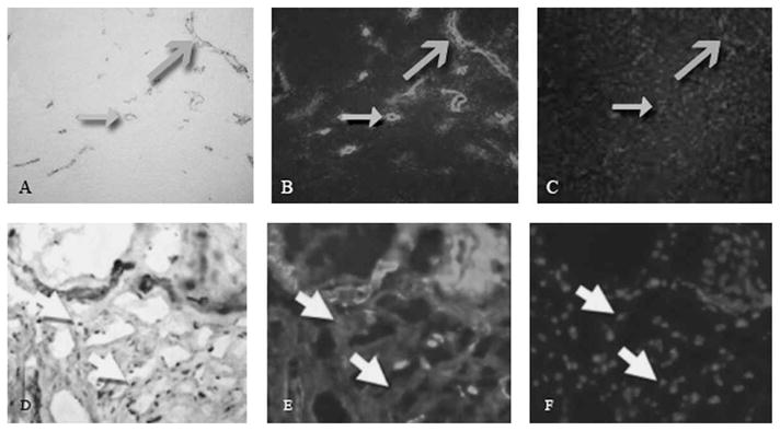
Fig 2.

A–F. Immunohistochemical analyses were done of the posterior joint capsule from an organ donor free of contractures (A–C), and a patient with a contracture (D–F). The images are from the same area of the same section for each specimen. Images A and D represent α-SMA (α-SMA antibody (original magnification, ×200). Images B and E represent laminin (Stain, laminin antibody; original magnification, ×200). Images C and F represent cell nuclei (Stain, DAPI; original magnification ×200). The closed grey arrows indicate the vascular structures in cross section, and the open grey arrows indicate the vascular structures in longitudinal section. The white arrows show areas where there is positive α-SMA staining without laminin indicating myofibroblasts. Myofibroblasts were seen in the contracture group, but were not seen in the control group.
