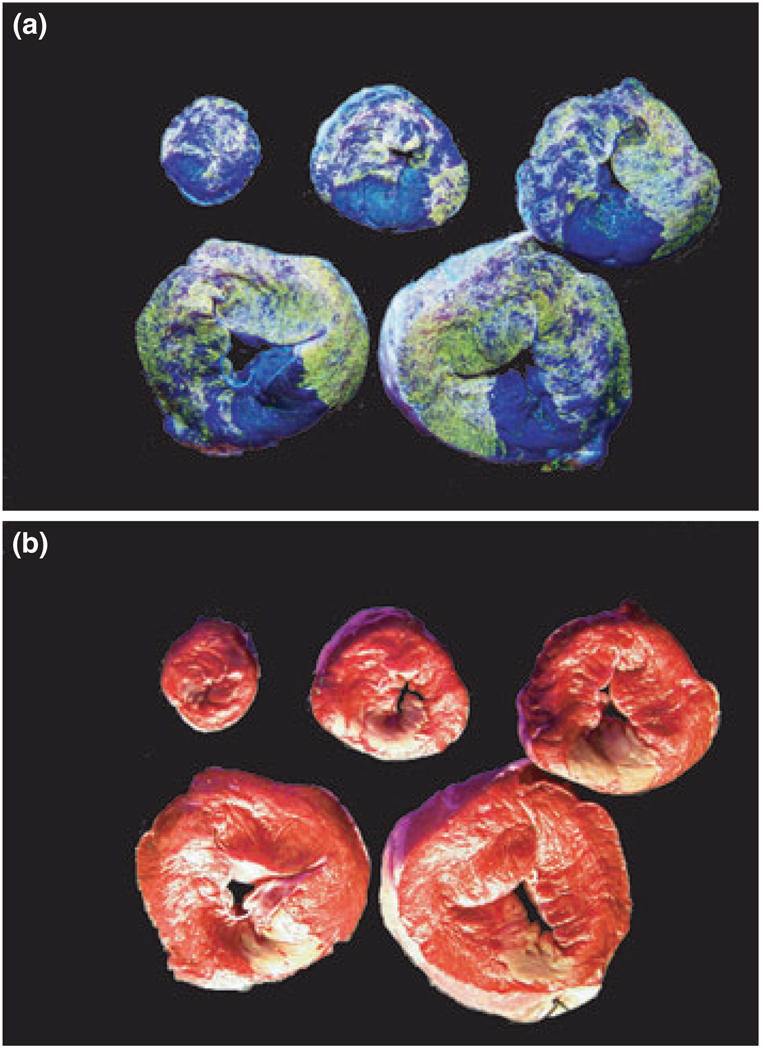
Figure 1.
Left ventricle slices infused with fluorescent microspheres and stained with triphenyl tetrazolium chloride (TTC) showing the area-at-risk and infarct area. Under ultraviolet light, area-at-risk were delineated by fluorescence-negativity (a) and infarct area were red-negative under full spectrum light (b).

