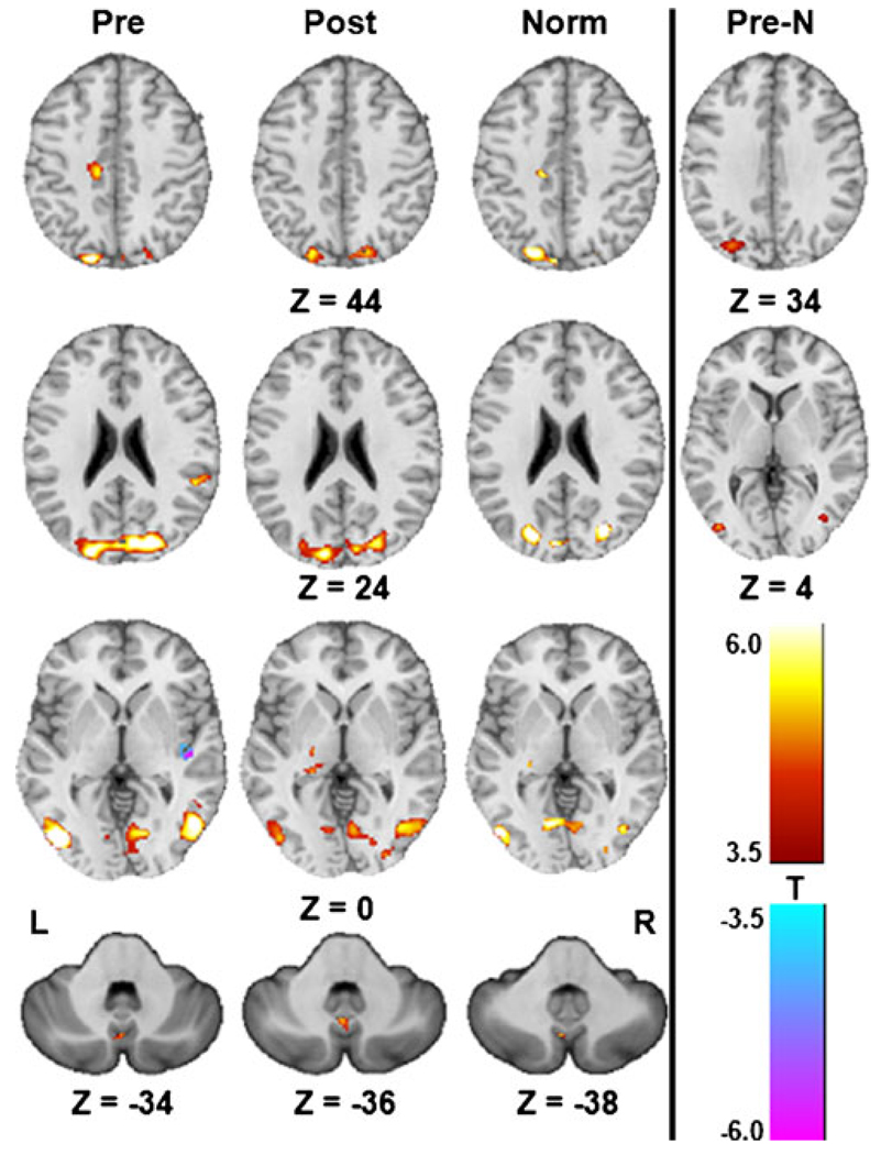
Fig. 5.
Activation patterns of the CBrot–CBstat contrast from the one-sample t-tests (Pre: pre-CN-NINM, Post: post-CN-NINM, Norm: normal controls) and the two-sample t-test comparing the pre-CN-NINM group to normal controls (Pre-N). All images are thresholded at α≤0.001 (|T11|≥4.0 for Pre and Post, |T8| ≥ 4.5 for Norm, and |T20|≥3.5 for Pre-N). Only clusters with a volume greater than 496 µl (128 µl for subcortical structures) are displayed. All Z values are in MNI coordinates

