
Figure 8.
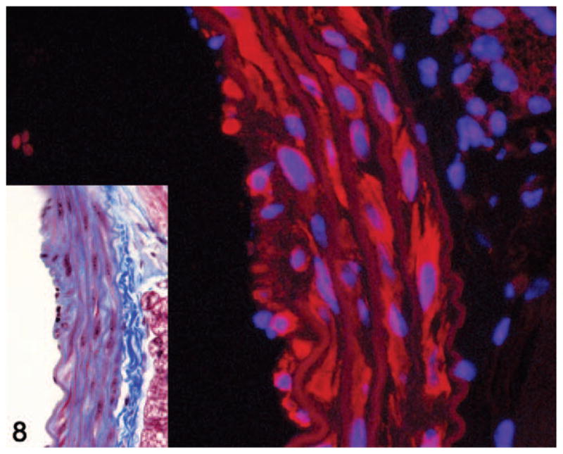
Aorta. Smoothelin immunofluorescence of a grade 2 lesion showing smoothelin-positive (red staining) and smoothelin-negative spindle cells within the media. Alexa Fluor 594 conjugate with DAPI counterstain. Inset shows histology of the same lesion. Masson’s trichrome.