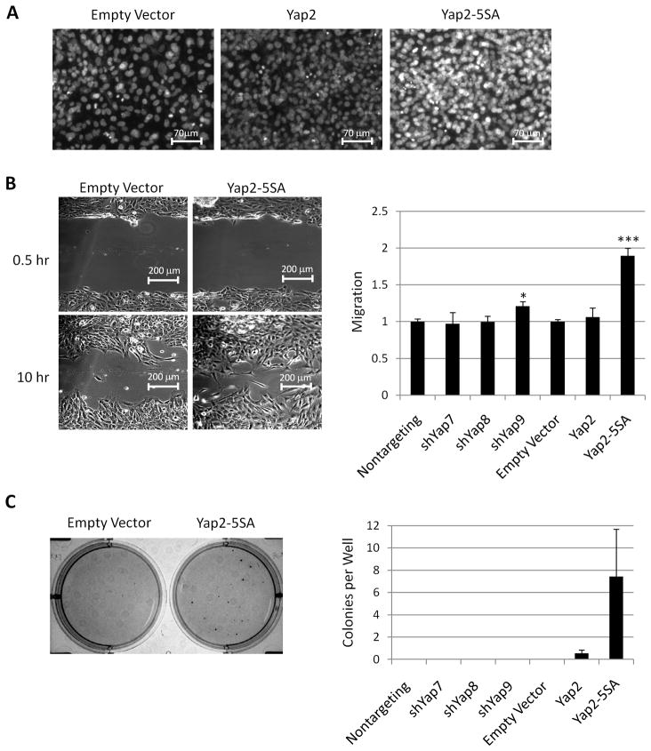
Figure 4.
Yap overexpression reduces contact inhibition and promotes wound closure and anchorage independent growth. A. Micrographs of confluent cultures stained for DNA to reveal cell density. Greater fluorescence intensity reveals multilayering of cells. B. Representative bright field micrographs (left) of control and Yap2-5SA cells 0.5 and 10 hours after wounding. Quantification of wound closure (right) after 10 hours. C. Representative micrograph of cell colonies visualized with MTT (left) after 14 days of growth in soft agar. Quantification of the number of colonies per well (right).*p<.05 and ***p<.001.

