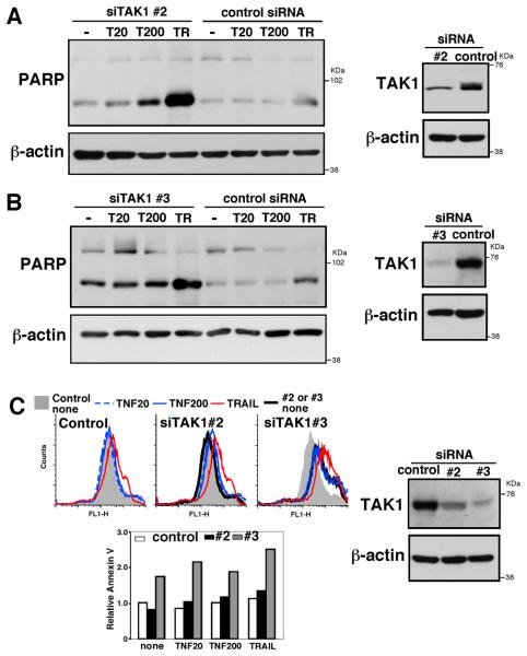
Fig. 5. Ablation of TAK1 in H-Ras transformed cells causes cell death.
(A, B) T24 cells were transfected with non-target siRNA (control) or TAK1 targeted siRNAs (siTAK1#2 (A) and siTAK1#3 (B)), and stimulated with 20 ng/ml TNF (T20), 200 ng/ml TNF (T200) for 4.5 h (A) or 10 h (B) or 100 ng/ml TRAIL (TR) for 4.5 h (A) or 2 h (B). Cell lysates were analyzed by immunoblotting with anti-TAK1 and anti-PAPR. (C) T24 cells were transfected with non-target, siTAK#2 or siTAK1#3, and stimulated with 20 ng/ml TNF (T20), 200 ng/ml TNF (T200) for 10 h or 100 ng/ml TRAIL (TR) for 2 h. Apoptotic cells were analyzed by Annexin V-binding assay. The staining of unstimulated non-target siRNA cells are shown in gray shadow all three panels (upper left panels). The fluorescence units relative to that of unstimulated non-target siRNA are shown (lower left panel). TAK1 expression was analyzed by immunoblotting with anti-TAK1 (right panels). Data are representative of two independent experiments with similar results.

