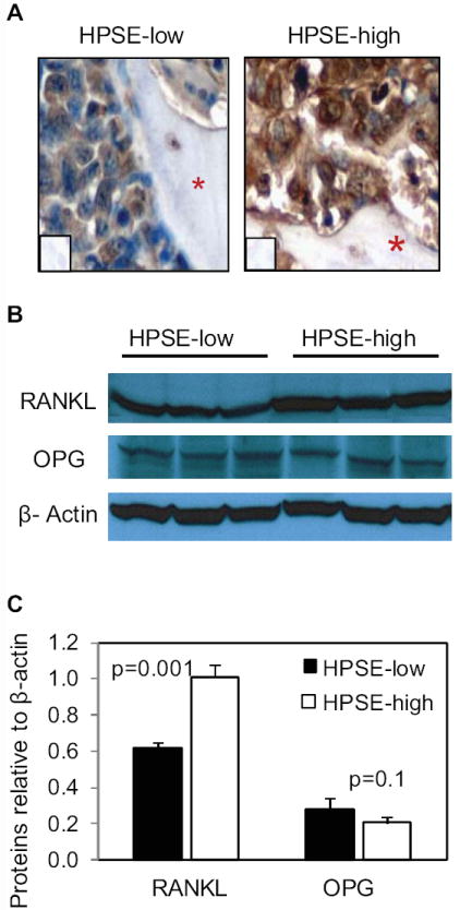
Figure 3. RANKL expression is elevated in tumors formed by HPSE-high cells.

A. Human RANKL staining within human bones from SCID-hu mice bearing tumors formed by HPSE-low or HPSE-high cells. The red asterisks denote bone and inserts are controls lacking addition of primary antibody (original magnification 400x). B. Primary myeloma tumors formed by HPSE-low or HPSE-high cells growing subcutaneously in SCID mice were extracted with detergent and western blotting was performed for detection of RANKL, OPG, and β-actin. Each lane of the blot represents tumor from a different animal. C. The bands from western blots were quantified relative to β-actin using ImageJ software. The bars represent means ± SD.
