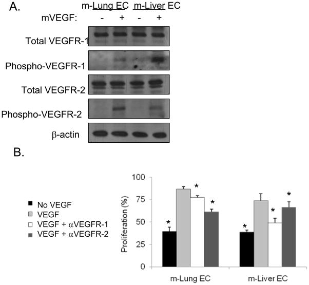
Figure 3. VEGF receptors on isolated mouse lung and liver ECs.
A, Western blot analysis for total and phospho-VEGFR-1 and VEGFR-2. Recombinant murine VEGF (mVEGF) was added where indicated. β-actin blot serves as loading control. B, EC proliferation (using BrdU incorporation). VEGF, αVEGFR-1, and/or αVEGFR-2 were added where indicated. All data are represented as a percentage with the addition of VEGF alone given a value of 100%. Bars represent standard deviation. *p<0.05 compared to VEGF group.

