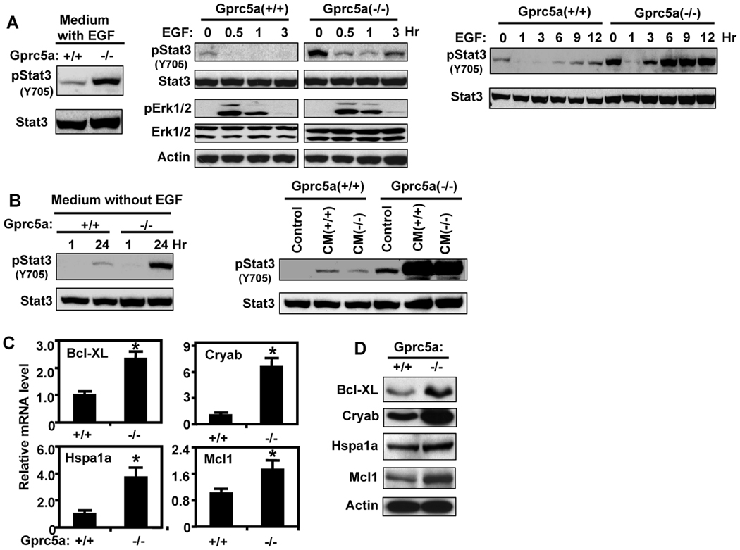
Figure 2.
EGF-independent Stat3 activation in Gprc5a−/− cells. A, left, Gprc5a+/+ and Gprc5a−/− cells were incubated in K-SFM with BPE and EGF (5 ng/ml) for 24 hours and then extracted and analyzed by immunoblotting. Middle, Gprc5a+/+ and Gprc5a−/− cells were starved overnight in K-SFM then treated with EGF (10 ng/ml) in K-SFM for up to 3 hours, then harvested and subjected to immunoblotting. Right, cells were starved overnight in K-SFM, treated with EGF (10 ng/ml) for up to 12 hours, and analyzed by immunoblotting. This treatment was done by replacing the basal culture medium with EGF containing medium at different times and harvesting the cells at the same time. The 0 hour point represents cells cultured in medium without EGF for 12 hours. The cells were harvested and subjected to immunoblotting. B, left, Gprc5a+/+ and Gprc5a−/− cells were cultured in K-SFM and harvested after 24 hours. In parallel cultures, the cells were cultured for 23 hours and then their “old” medium was replaced with fresh medium and the cells were incubated in this medium for one hour. The cells were harvested and analyzed by immunoblotting. Right, Gprc5a+/+ and Gprc5a−/− cells were incubated in medium conditioned for 24 hours by other cultures of either Gprc5a+/+ or Gprc5a−/− cells as indicated. The cells were harvested after one hour and analyzed by immunoblotting. C, Gprc5a+/+ and Gprc5a−/− cells were starved for 48 hours and extracted for mRNA analysis by QPCR for Stat3 regulated genes. *, P < 0.05. D, cells treated as in C were analyzed by western blotting for Stat3 regulated proteins. Actin was used to control for loading.

