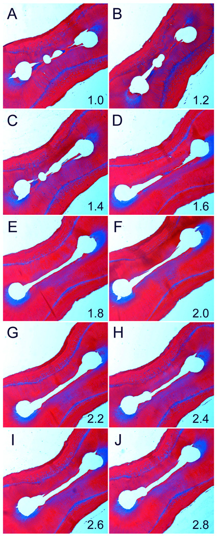
Fig.6.
A representative example of a root specimen that was cleaned using the ANP technique. Figures 6A to 6J represent Masson’s trichrome-stained sections of the same root taken at the ten canal levels. No debris could be detected at each canal level. The number at the lower right corner of each image represents canal level in mm (Original magnification 20–4×).

