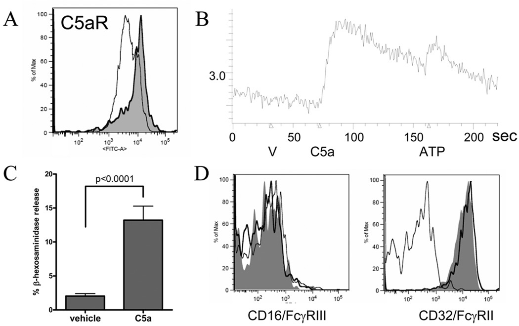
Figure 5.
A. Human SMC were stained with anti-C5aR (shaded) or isotype and assessed by flow cytometry. C3aR was not detectable (data not shown). B. Ca flux in SMC (V=vehicle, C5a=C5a 100nm, ATP=adensine triphosphate positive control). A and B representative of 2 experiments using distinct SMC cultures. C. Degranulation in SMC treated with C5a 10nM for 4–6h, pooled from 7 experiments (n=14/group). Degranulation was observed with as little as 1nM C5a and did not increase measurably from 10–300nM (data not shown). D. SMC stained for FcγR expression 2h after incubation with C5a 100nM (shaded) or vehicle (dark line) compared to isotype (light line). FcγRI (CD64) remained absent, as did 2B6 staining for FcγRIIB (data not shown). Data confirmed in 3 SMC cultures in 2 experiments.

