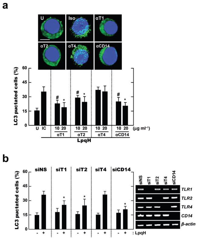
Fig. 2. TLR2/1 and CD14, but not TLR4, are required for LpqH-induced autophagy.
A. Human primary monocytes were incubated with LpqH in the absence or presence of neutralizing Ab for hTLR1 (αT1), hTLR2 (αT2), hTLR4 (αT4), hCD14 (αCD14) or isotype control (IC; mIgG1 and mIgG2a; shown is the data for mIgG1 Ab), and subjected to confocal analysis as described in Fig. 1A. Top, a representative image with similar results is shown (three experiments); bottom, quantification of data; LC3 punctated cells were counted manually in DAPI-stained monocytes. #P < 0.02; *P < 0.001, versus control condition. Scale bars, 5 μm.
B. Human THP-1 cells transfected with non-specific siRNA (siNS) or specific siRNAs for hTLR1 (siT1), hTLR2 (siT2), hTLR4 (siT4) or hCD14 (siCD14) were incubated in the absence or presence of LpqH (100 ng ml−1) for 24 h, and subjected to confocal analysis as described in Fig. 1A. Graph is shown the percentage of LC3 punctated cells; right, RT-PCR for transfection efficiency. *P < 0.008, versus control condition. Data shown (for A and B) represent the means ± SD of three independent samples, with each experiment including at least 250 cells scored in five random fields. U, untreated and incubated.

