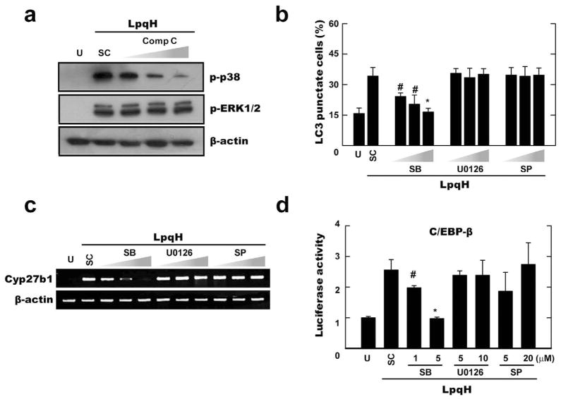
Fig. 6. AMPK-p38 MAPK pathway plays a role for autophagy activation and C/EBP-β-dependent Cyp27b1 hydroxylase induction in response to LpqH stimulation.
A–C. Human monocytes were stimulated with LpqH (100 ng ml−1) in the absence or presence of various inhibitors (AMPK inhibitor compound C; Comp C, 5, 10, 25 μM; for A) or MAPK inhibitors [p38, SB203580 (SB), 1, 5, 10 μM; ERK, U0126, 5, 10, 20 μM; JNK, SP600125 (SP), 5, 10, 20 μM; for B and C], and then subjected to Western blot (for A), confocal analysis (for B), or semi-quantitative RT-PCR analysis (for C). (B) Data shown represent the means ± SD of three independent samples, with each experiment including at least 250 cells scored in five random fields. (A and C) Representative images are shown (at least three independent experiments).
D. THP-1 cells were transfected with a C/EBP-β luciferase reporter construct. After 36 h, cells were pre-treated with MAPK inhibitors (the same conditions as shown in C), followed by LpqH stimulation for 6 h. The luciferase activity was then measured and normalized to β-galactosidase activity.
#P < 0.01; *P < 0.0004, versus control conditions. U, untreated and incubated; SC, treated with solvent control (0.1% DMSO).

