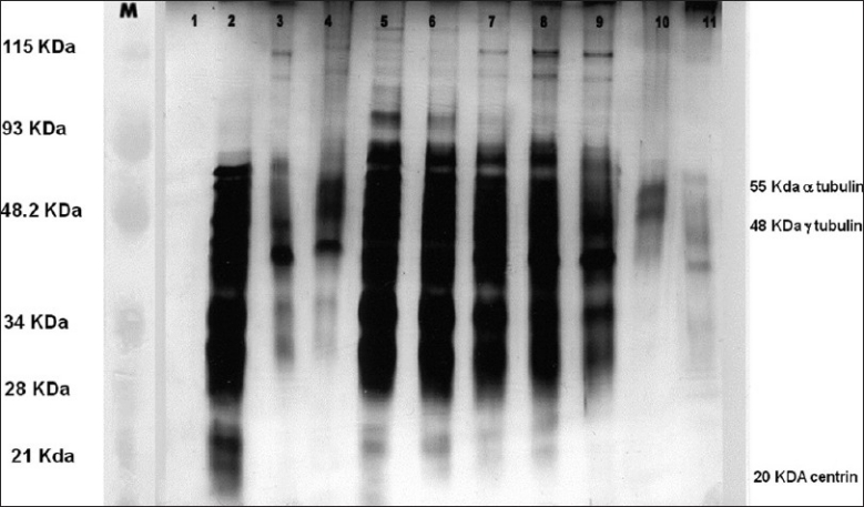
Figure 1.

Silver staining of isolated sperm centrosomal fractions on a 12% SDS-PAGE. Lanes 1 to 11 - The various fractions collected by discontinuous sucrose density gradient method. Fractions 2-9 contains maximum proteins i.e., centrin, α and γ tubulin. Lane M - Broad range rainbow protein molecular weight marker (12,000 - 2,25,000, Amersham Biosciences now GE Healthcare Lifesciences)
