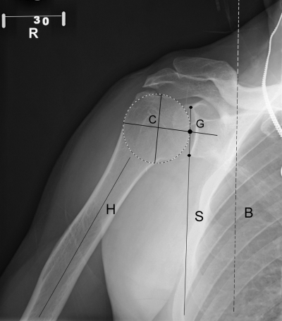
Fig. 1.
Radiographic measurement of glenohumeral kinematics. The geometric center of the humeral head (C) was located first with a so-called best-fit circle positioned over the humeral head outline. The superior and inferior rims of the glenoid were then marked to demarcate the glenoid line. The glenoid center point (G) was then located automatically by the software. The vertical distance from the geometric center of the humeral head to the glenoid center was measured for superior humeral head migration. The angle formed by the line drawn along the long axis of the humerus (H) and the glenoid line (S) was measured to calculate the glenohumeral angle. The glenoid line (S) was compared with the vertical axis of the body (B) to calculate the scapulothoracic angle. A reference bar (R) with a known length (5 cm) was always included in radiographs to adjust for magnification differences.

