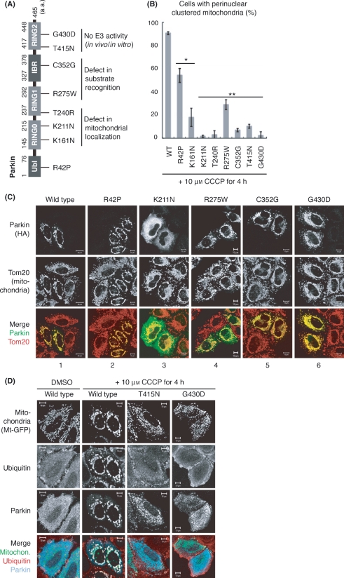
Figure 3.
Various pathogenic mutations of Parkin impede mitochondrial clustering. (A) Schematic diagram of disease-relevant mutants of Parkin used in this study. IBR, in between RING; Ubl, ubiquitin like. (B) HeLa cells expressing HA-Parkin with various pathogenic mutations were treated with CCCP followed by immunocytochemistry. Mitochondrial clustering was analyzed in more than 100 cells per mutation. Bars represent the mean ± SD values of at least three experiments. Asterisk, P < 0.01; two asterisks, P < 0.001 (Welch’s t-test). (C) Immunocytochemistry indicative of a typical example for each mutation is shown. Scale bars represent 20 μm in wild type and 10 μm in the other mutations. (D) Triple-staining using mitochondria-targeting GFP (Mt-GFP), anti-ubiquitin and anti-Parkin antibodies confirmed that mitochondria in T415N and G430D mutants did not undergo ubiquitylation or clustering.

