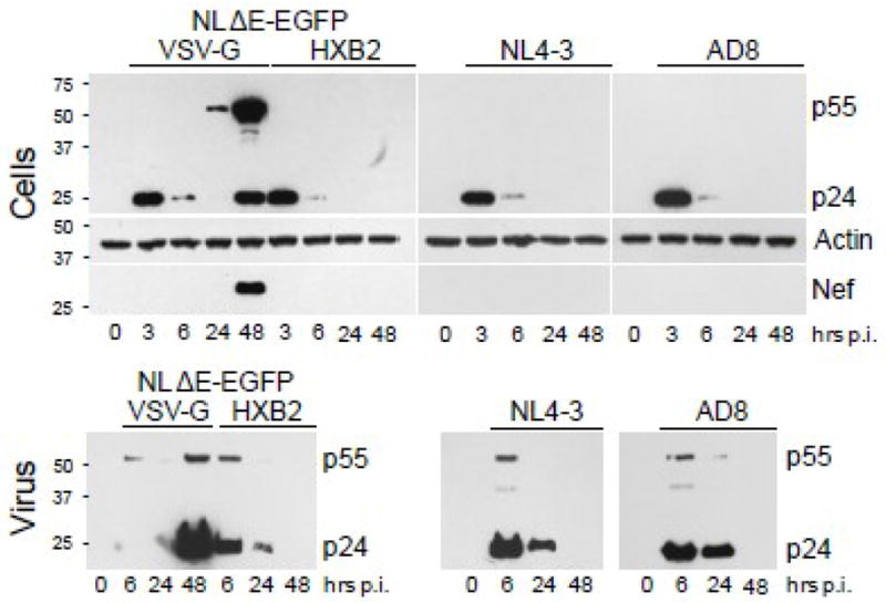
Fig. 4. Immunoblot analysis of HIV-1 proteins expressed in podocytes and released virions.

Podocytes were infected for 3 h with NL4-3, AD8 or NLΔE-EGFP pseudotyped with HXB2 or VSV-G envelopes. Cellular lysates (Cells) were prepared at indicated times and analyzed by Western blotting for the expression of HIV-1 Gag (p24, p55), Nef, and actin. After 3 h infection, the cells were washed 5 times and virus particles released from infected cells were sedimented from the same volume of media (6 ml) collected between 3 h and 6 h (denoted as 6 h), between 6 h and 24 h (24 h), and between 24 h and 48 h post-infection (48 h) (Virus). Pelleted virions were analyzed by immunoblotting for the expression of HIV-1 Gag.
