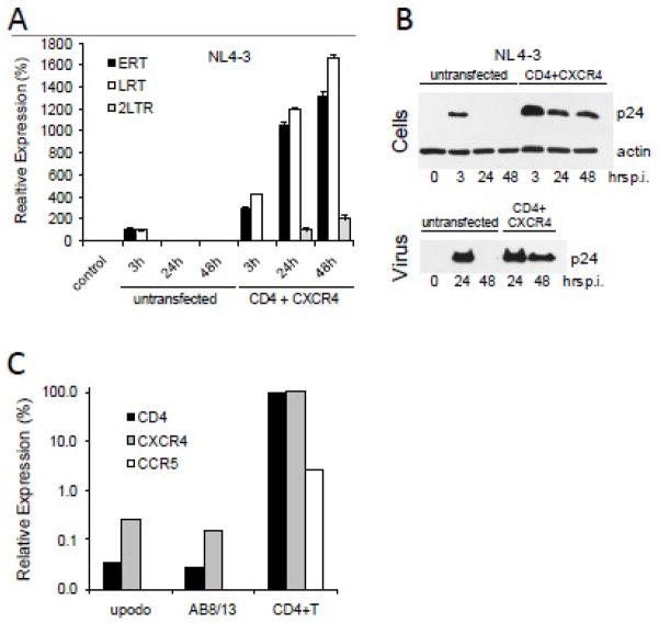
Fig. 6. Productive infection of podocytes expressing CD4 and CXCR4 receptors.
(A) Undifferentiated AB8/13 podocytes were transfected with CD4 and CXCR4 expression vectors using PolyFect (Qiagen). Twenty-four hours later, the cells were infected with NL4-3 for 3 h. Cellular DNA was analyzed by real-time PCR for the expression of ERT, LRT and 2-LTR circles. Expression was normalized against GAPDH. Relative expression levels were compared by individually setting the levels of ERT and LRT at 3 h post-infection of untransfected cells to 100%. Uninfected cells (control) show undetectable levels of HIV-1 DNA. (B) Lysates from infected podocytes (Cells) and pelleted virions (Virus), prepared as described in Fig. 4 legend, were analyzed for the expression of HIV-1 Gag p24. Time “0” represents uninfected cells. (C) Differentiated AB8/13 podocytes and primary urinary podocytes (upodo) show negligible expression of CD4, CXCR4 and CCR5 mRNAs. Human blood-derived PHA/IL-2 activated CD4+ T cells (Khatua et al., 2009) express CD4, CXCR4 and CCR5 mRNAs and served as a positive control.

