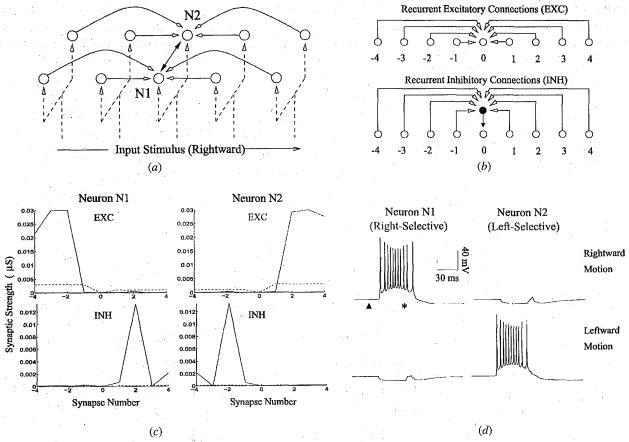
Fig 3.
Detecting Multiple Directions of Motion. (a) A model network consisting of two chain of recurrently connected neurons receiving retinotopic inputs. A given neuron receives recurrent excitation and recurrent inhibition(white-headed arrows) as well as inhibition (darkheaded arrows) from its counterpart in the other chain. (b) Recurrent connections to a given neuron (labeled ‘0’) arise from 4 preceding and 4 succeeding neurons in its chain. Inhibition at a given neuron is mediated via a GABAergic interneuron (darkened circle). (c) Synaptic strength of recurrent excitatory (EXC) and inhibitory (INH) connections to neurons N1 and N2 before (dotted lines) and after learning (solid lines). Synapses were adapted during 100 trials of exposure to alternating leftward and rightward moving stimuli. (d) Responses of neurons N1 and N2 to rightward and leftward moving stimuli. After learning, neuron N1 has become selective for rightward motion (as have other neurons in the same chain) while neuron N2 has become selective for leftward motion. In the preferred direction, each neuron starts firing several milliseconds before the input arrives at its soma (marked by an asterisk) due to recurrent excitation from preceding neurons. The dark triangle represents the start of input stimulation in the network.

