

Abstract
In the title compound, C6H3F3, weak electrostatic and dispersive forces between C(δ+)—F(δ−) and H(δ+)—C(δ−) groups are at the borderline of the hydrogen-bond phenomenon and are poorly directional and further deformed in the presence of π–π stacking interactions. The molecule lies on a twofold rotation axis. In the crystal structure, one-dimensional tapes are formed via two antidromic C—H⋯F hydrogen bonds. These tapes are, in turn, connected into corrugated two-dimensional sheets by bifurcated C—H⋯F hydrogen bonds. Packing in the third dimension is furnished by π–π stacking interactions with a centroid–centroid distance of 3.6362 (14) Å.
Related literature
For C—H⋯F interactions, see: Althoff et al. (2006 ▶); Bats et al. (2000 ▶); Choudhury et al. (2004 ▶); D’Oria & Novoa (2008 ▶); Dunitz & Taylor (1997 ▶); Howard et al. (1996 ▶); Müller et al. (2007 ▶); O’Hagan (2008 ▶); Reichenbacher et al. (2005 ▶); Weiss et al. (1997 ▶). For related crystal structures of several polyfluorinated benzenes, see: Thalladi et al. (1998 ▶). For crystallization techniques, see: Boese & Nussbaumer (1994 ▶).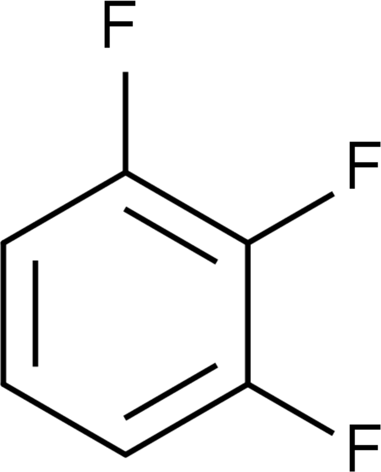
Experimental
Crystal data
C6H3F3
M r = 132.08
Monoclinic,

a = 7.4238 (19) Å
b = 11.590 (3) Å
c = 7.0473 (17) Å
β = 112.783 (4)°
V = 559.1 (2) Å3
Z = 4
Mo Kα radiation
μ = 0.16 mm−1
T = 233 K
0.30 × 0.30 × 0.30 mm
Data collection
Siemens SMART three-axis goniometer with an APEXII area-detector system diffractometer
Absorption correction: multi-scan (SADABS; Bruker; 2004 ▶) T min = 0.820, T max = 0.953
1074 measured reflections
634 independent reflections
413 reflections with I > 2σ(I)
R int = 0.013
Refinement
R[F 2 > 2σ(F 2)] = 0.061
wR(F 2) = 0.226
S = 1.04
634 reflections
44 parameters
H-atom parameters constrained
Δρmax = 0.20 e Å−3
Δρmin = −0.18 e Å−3
Data collection: APEX2 (Bruker, 2008 ▶); cell refinement: SAINT (Bruker, 2008 ▶); data reduction: SAINT; program(s) used to solve structure: SHELXTL (Sheldrick, 2008); program(s) used to refine structure: SHELXTL; molecular graphics: Mercury (Macrae et al., 2008 ▶) and GIMP2 (The GIMP team, 2008 ▶); software used to prepare material for publication: publCIF (Westrip, 2009 ▶).
Supplementary Material
Crystal structure: contains datablocks I, global. DOI: 10.1107/S1600536809038975/lh2880sup1.cif
Structure factors: contains datablocks I. DOI: 10.1107/S1600536809038975/lh2880Isup2.hkl
Additional supplementary materials: crystallographic information; 3D view; checkCIF report
Table 1. Hydrogen-bond geometry (Å, °).
| D—H⋯A | D—H | H⋯A | D⋯A | D—H⋯A |
|---|---|---|---|---|
| C3—H3⋯F2i | 1.10 | 2.77 | 3.560 (3) | 129 |
| C3—H3⋯F1ii | 1.10 | 2.59 | 3.528 (4) | 144 |
| C4—H4⋯F2iii | 1.00 | 2.60 | 3.440 (4) | 142 |
Symmetry codes: (i)  ; (ii)
  ; (iii)
  .
.
Acknowledgments
MTK and RB thank the DFG FOR-618. GRD thanks the DST for the award of a J.C. Bose fellowship. TST thanks the UGC for an SRF.
supplementary crystallographic information
Comment
Despite the high electronegativity difference between carbon and fluorine, the C–F bond acts as a poor hydrogen bond acceptor due to the hardness of the F-atom (Dunitz & Taylor, 1997; O'Hagan, 2008). The resultant weak C–H···F–C interactions (Howard et al., 1996; Reichenbacher et al., 2005) arise mainly due to electrostatic and dispersive forces between the C(δ+)–F(δ-) and the H(δ+)–C(δ-) fragments. These interactions, at the borderline of the hydrogen bond phenomenon, are also poorly directional and are deformed by other dominant interactions (Weiss, et al., 1997; D'Oria & Novoa, 2008; Müller et al., 2007). In the absence of other interactions these weak interactions can play a role in the overall crystal packing of the molecule (Bats et al. 2000; Choudhury et al. 2004; Althoff et al. 2006). In activated systems such as polyfluorobenzenes, C–H···F–C interactions may be of significance, and in connection there are some reports of the crystal structures of several polyfluorinated benzene compunds (Thalladi et al., 1998). As a continuation of this work, we report here the crystal structure 1,2,3-trifluorobenzene (1). The comparison crystal structures of 1,2- and 1,4-difluorobenzene and 1,3,5-trifluorobenzene have been reported in this earlier work.
Experimental
The crystals were prepared from commerical samples by zone melting in a quartz capillary at 235 K (1) according to the procedure outlined by (Boese & Nussbaumer, 1994).
Refinement
Treatment of hydrogen atoms: Riding model with the 1.2 fold isotropic displacement parameters of the equivalent Uij of the corresponding carbon atom.
Figures
Fig. 1.

Part of the crystal structure of 1 (a) 2D network of C–H···F–C interactions viewed along the c-axis (b) π–π stacking of molecules viewed along the c-axis.
Fig. 2.

The molecular structure of (1) with displacement ellipsoids drawn at the 50% probability level. The identically labelled atoms are related to each other by the symmetry operator (2-x, y, -z+1/2).
Crystal data
| C6H3F3 | F(000) = 264 |
| Mr = 132.08 | Dx = 1.569 Mg m−3 |
| Monoclinic, C2/c | Mo Kα radiation, λ = 0.71073 Å |
| Hall symbol: -C 2yc | Cell parameters from 376 reflections |
| a = 7.4238 (19) Å | θ = 3.8–22.7° |
| b = 11.590 (3) Å | µ = 0.16 mm−1 |
| c = 7.0473 (17) Å | T = 233 K |
| β = 112.783 (4)° | Cylindric, colourless |
| V = 559.1 (2) Å3 | 0.30 × 0.30 × 0.30 mm |
| Z = 4 |
Data collection
| Siemens SMART three-axis goniometer with an APEXII area-detector system diffractometer | 634 independent reflections |
| Radiation source: fine-focus sealed tube | 413 reflections with I > 2σ(I) |
| graphite | Rint = 0.013 |
| Detector resolution: 512 pixels mm-1 | θmax = 28.2°, θmin = 3.5° |
| in ω at 0.3° scan width one run with 740 frames, phi = 0°, chi = 0° | h = −9→9 |
| Absorption correction: multi-scan (SADABS; Bruker; 2004) | k = −14→10 |
| Tmin = 0.820, Tmax = 0.953 | l = −9→4 |
| 1074 measured reflections |
Refinement
| Refinement on F2 | Primary atom site location: structure-invariant direct methods |
| Least-squares matrix: full | Secondary atom site location: difference Fourier map |
| R[F2 > 2σ(F2)] = 0.061 | Hydrogen site location: inferred from neighbouring sites |
| wR(F2) = 0.226 | H-atom parameters constrained |
| S = 1.04 | w = 1/[σ2(Fo2) + (0.1501P)2 + 0.039P], where P = (Fo2 + 2Fc2)/3 |
| 634 reflections | (Δ/σ)max = 0.017 |
| 44 parameters | Δρmax = 0.20 e Å−3 |
| 0 restraints | Δρmin = −0.18 e Å−3 |
Special details
| Geometry. All e.s.d.'s (except the e.s.d. in the dihedral angle between two l.s. planes) are estimated using the full covariance matrix. The cell e.s.d.'s are taken into account individually in the estimation of e.s.d.'s in distances, angles and torsion angles; correlations between e.s.d.'s in cell parameters are only used when they are defined by crystal symmetry. An approximate (isotropic) treatment of cell e.s.d.'s is used for estimating e.s.d.'s involving l.s. planes. |
| Refinement. Refinement of F^2^ against ALL reflections. The weighted R-factor wR and goodness of fit S are based on F^2^, conventional R-factors R are based on F, with F set to zero for negative F^2^. The threshold expression of F^2^ > σ(F^2^) is used only for calculating R-factors(gt) etc. and is not relevant to the choice of reflections for refinement. R-factors based on F^2^ are statistically about twice as large as those based on F, and R- factors based on ALL data will be even larger. |
Fractional atomic coordinates and isotropic or equivalent isotropic displacement parameters (Å2)
| x | y | z | Uiso*/Ueq | ||
| F1 | 1.0000 | 0.30558 (17) | 0.2500 | 0.1156 (10) | |
| F2 | 0.6666 (2) | 0.4183 (2) | 0.1576 (3) | 0.1354 (10) | |
| C1 | 1.0000 | 0.4213 (3) | 0.2500 | 0.0769 (9) | |
| C2 | 0.8308 (3) | 0.4803 (2) | 0.2036 (3) | 0.0824 (8) | |
| C3 | 0.8265 (4) | 0.5973 (3) | 0.2023 (3) | 0.0942 (9) | |
| H3 | 0.6833 | 0.6388 | 0.1585 | 0.113* | |
| C4 | 1.0000 | 0.6558 (3) | 0.2500 | 0.1006 (13) | |
| H4 | 1.0000 | 0.7422 | 0.2500 | 0.121* |
Atomic displacement parameters (Å2)
| U11 | U22 | U33 | U12 | U13 | U23 | |
| F1 | 0.161 (2) | 0.0623 (13) | 0.1249 (16) | 0.000 | 0.0563 (14) | 0.000 |
| F2 | 0.0959 (12) | 0.157 (2) | 0.1484 (16) | −0.0341 (10) | 0.0415 (10) | 0.0067 (12) |
| C1 | 0.1030 (19) | 0.0573 (16) | 0.0725 (15) | 0.000 | 0.0364 (13) | 0.000 |
| C2 | 0.0830 (14) | 0.0890 (17) | 0.0770 (13) | −0.0101 (9) | 0.0327 (10) | 0.0013 (9) |
| C3 | 0.1073 (17) | 0.0935 (18) | 0.0858 (15) | 0.0277 (12) | 0.0419 (12) | 0.0094 (10) |
| C4 | 0.163 (4) | 0.0605 (17) | 0.0848 (19) | 0.000 | 0.056 (2) | 0.000 |
Geometric parameters (Å, °)
| F1—C1 | 1.341 (4) | C3—C4 | 1.377 (3) |
| F2—C2 | 1.342 (3) | C3—H3 | 1.0973 |
| C1—C2 | 1.354 (3) | C4—H4 | 1.0018 |
| C2—C3 | 1.357 (4) | ||
| F1—C1—C2 | 120.30 (15) | C2—C3—H3 | 117.3 |
| C2i—C1—C2 | 119.4 (3) | C4—C3—H3 | 124.4 |
| F2—C2—C3 | 121.1 (2) | C3—C4—C3i | 121.0 (3) |
| F2—C2—C1 | 117.3 (3) | C3—C4—H4 | 119.5 |
| C3—C2—C1 | 121.5 (2) | C3i—C4—H4 | 119.5 |
| C2—C3—C4 | 118.3 (2) |
Symmetry codes: (i) −x+2, y, −z+1/2.
Hydrogen-bond geometry (Å, °)
| D—H···A | D—H | H···A | D···A | D—H···A |
| C3—H3···F2ii | 1.10 | 2.77 | 3.560 (3) | 129 |
| C3—H3···F1iii | 1.10 | 2.59 | 3.528 (4) | 144 |
| C4—H4···F2iv | 1.00 | 2.60 | 3.440 (4) | 142 |
Symmetry codes: (ii) −x+1, −y+1, −z; (iii) x−1/2, y+1/2, z; (iv) x+1/2, y+1/2, z.
Footnotes
Supplementary data and figures for this paper are available from the IUCr electronic archives (Reference: LH2880).
References
- Althoff, G., Ruiz, J., Rodriguez, V., Lopez, G., Perez, J. & Janiak, C. (2006). CrystEngComm, 8, 662–665.
- Bats, J. W., Parsch, J. & Engels, J. W. (2000). Acta Cryst. C56, 201–205. [DOI] [PubMed]
- Boese, R. & Nussbaumer, M. (1994). In Situ Crystallisation Techniques In Organic Crystal Chemistry, edited by D. W. Jones, pp. 20–37. Oxford University Press.
- Bruker (2004). SADABS Bruker AXS Inc., Madison, Wisconsin, USA.
- Bruker (2008). APEX2 and SAINT Bruker AXS Inc., Madison, Wisconsin, USA.
- Choudhury, A. R., Nagarajan, K. & Guru Row, T. N. (2004). Acta Cryst. C60, o644–o647. [DOI] [PubMed]
- D’Oria, E. & Novoa, J. J. (2008). CrystEngComm, 10, 423–436.
- Dunitz, J. D. & Taylor, R. (1997). Chem. Eur. J.3, 89–98.
- Howard, J. A. K., Hoy, V. J., O’Hagan, D. & Smith, G. T. (1996). Tetrahedron, 38, 12613–12622.
- Macrae, C. F., Bruno, I. J., Chisholm, J. A., Edgington, P. R., McCabe, P., Pidcock, E., Rodriguez-Monge, L., Taylor, R., van de Streek, J. & Wood, P. A. (2008). J. Appl. Cryst.41, 466–470.
- Müller, K., Faeh, C. & Diederich, F. (2007). Science, 317, 1881–1886. [DOI] [PubMed]
- O’Hagan, D. (2008). Chem. Soc. Rev.37, 308–319.
- Reichenbacher, K., Suss, H. I. & Hulliger, J. (2005). J. Chem. Soc. Rev.34, 22–30. [DOI] [PubMed]
- Thalladi, V. R., Weiss, H. C., Bläser, D., Boese, R., Nangia, A. & Desiraju, G. R. (1998). J. Am. Chem. Soc.120, 8702–8710.
- The GIMP team (2008). The GNU Image Manipulation Program, http://www.gimp.org.
- Weiss, H. C., Boese, R., Smith, H. L. & Haley, M. M. (1997). Chem. Commun. pp. 2403–2404.
- Westrip, S. P. (2009). publCIF. In preparation.
Associated Data
This section collects any data citations, data availability statements, or supplementary materials included in this article.
Supplementary Materials
Crystal structure: contains datablocks I, global. DOI: 10.1107/S1600536809038975/lh2880sup1.cif
Structure factors: contains datablocks I. DOI: 10.1107/S1600536809038975/lh2880Isup2.hkl
Additional supplementary materials: crystallographic information; 3D view; checkCIF report