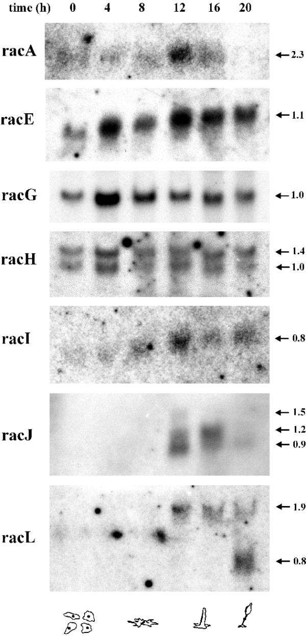
Figure 2.

Patterns of mRNA expression of rac genes during the developmental cycle. Dictyostelium cells were allowed to develop on nitrocellulose filters and total RNA was extracted at the indicated time points. Northern blots containing 20 µg total RNA for each time point were hybridised with the corresponding 32P-radiolabelled probe. Two different blots were used; for racA, racE, racI, racJ and racL the same blot was used; for racG and racH a separate blot was used. The lane corresponding to 4 h was slightly overloaded. The blots were stripped and tested for absence of radioactive signals prior to hybridisation with a new probe. The arrows mark the size of the transcripts in kilobases. The developmental stages corresponding to the time points are depicted below. The transcripts of some of the rac genes analysed here, most notably racJ and racL, are present after the aggregation stage. Very long exposure times were required in order to detect racA, racI, racJ and racL.
