Figure 7.
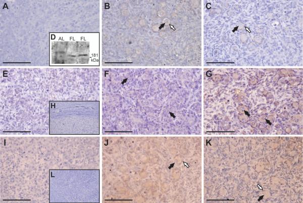
Expression and localisation of ROBO1, ROBO2 and SLIT2 throughout gestation. A, Light-field microscopy of a Stage 1 ovary showing no positive brown DAB staining for ROBO1. Light-field microscopy of Stage 2 (B) and Stage 3 (C) ovaries showing ROBO1 staining around the pre-granulosa cells of developing primoridal follicles. At no point was the positive staining observed in the migrating cells. Black arrows are pointing towards the oocytes. The pre-granulosa cells are highlighted by white arrows. D, Immunoblot analysis of ROBO1 clarifying the specificity of the antibody. As expected, a 181 kDa protein, was detected in the total protein lysate from two fetal liver samples (FL) but not in a lamb liver sample (AL). E, Weak positive brown DAB staining for ROBO2 was detected in Stage 1 ovaries using light-field microscopy. F Some ROBO2 staining was noted in Stage 2 ovaries around germ cell clusters and early primordial follicles. G, The oocytes of developing primordial follicles showed intense positive brown staining in Stage 3 of gestation. Black arrows are pointing towards the oocytes. H, A negative control serial section contained no ROBO2 staining. I, No positive brown DAB staining for SLIT2 was noted in Stage 1 ovaries by light-field microscopy. J, Positive SLIT2 expression was observed in early primordial follicles of Stage 2 ovaries. K, Weak SLIT2 staining was detected in the oocytes and pre-granulosa cells of developing primordial follicles at Stage 3 of gestation. Black arrows are pointing towards oocytes. Pre-granulosa cells are highlighted by white arrows. L, A negative control section showed no SLIT2 staining. All the scale bars represent 100 μm.

