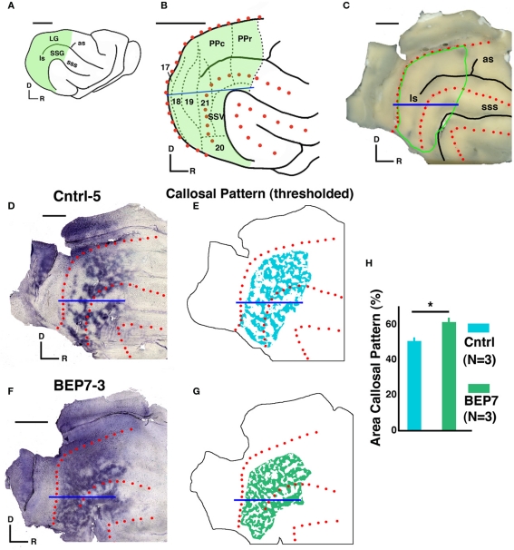
Figure 1.
Effect of bilateral enucleation on postnatal day 7 (BEP7) on the distribution of interhemispheric visual callosal connections in the ferret. The distribution of callosal connections in one hemisphere of adult BEP7 and control ferrets were studied following multiple intracortical injections of the tracer HRP in the contralateral hemisphere. Green areas in (A) and (B) include regions of visual cortex analyzed. Approximate locations of visual areas described in previous reports are indicated in (B); the blue line marks the representation of the horizontal meridian. Red dots indicate the crown of the suprasylvian and ectosylvian and gyri, and the dorsal/caudal edge of the occipital lobe, which were marked directly on the brains before flattening. (C) Flattened brain before sectioning, area outlined by green line contains visual areas analyzed. Labeled callosal connections (labeled somas and axon terminations) appear as dark areas in (D) and (F); and as colored areas in the thresholded versions (E, G). The percent area occupied by callosal connections was significantly (p < 0.05) greater in BEP7 ferrets than in Control ferrets (H). LG, lateral gyrus; PPc, posterior parietal caudal area; PPr, posterior parietal rostral area; SSG, suprasylvian gyrus; SSV, suprasylvian visual areas; as, ansate sulcus; ls, lateral sulcus, sss; suprasylvian sulcus. Scale = 5 mm.

