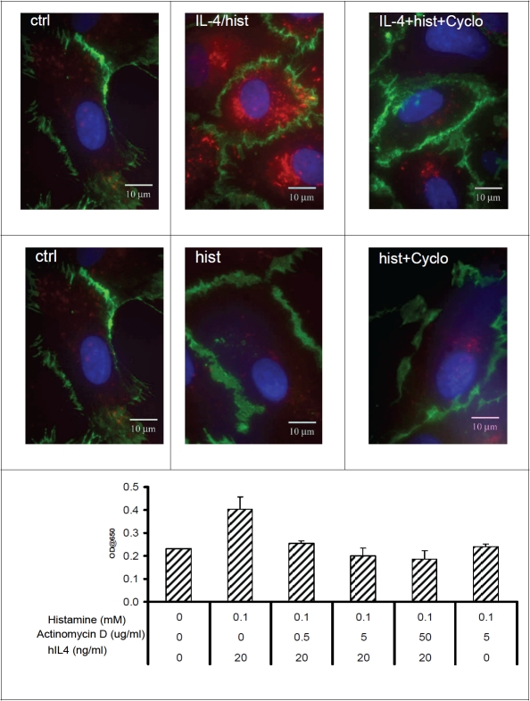
Figure 4.
hlL-4 induced expression of Psel requires protein synthesis: Representative figures from localization studies of Psel (red) and PECAM-1 (green) in HMVEC,c, treated with IL-4/histamine and histamine alone in presence or absence of cycloheximide. After treatment, the cells were permeabilized and double IF were done as described in material and methods. PECAM-1 is constitutively expressed on the lateral borders of the cell. Cell nuclei are stained with DAPI (blue). Lower panel showing dose-dependent inhibition of cell surface Psel induction by actinomycin D. Monolayer of HMVEC,c were stimulated with indicated IL-4 and/or histamine along with actinomycin D for 24 h. Cell surface Psel was measured by cell-ELISA as described in materials and methods. Data are from one experiment representative of at least three independent experiments per condition.

