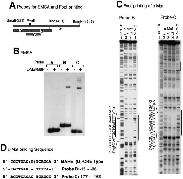
Figure 7.

Identification of c-Maf-binding sequences on the c-maf gene. (A) Schematic representation of probes used for EMSA and footprinting analyses. (B) EMSA of c-maf promoter probes with c-Maf–MBP protein. EMSA was carried out as in the legend to Figure 6 and Materials and Methods. (C) Footprinting analysis of the c-maf gene promoter with c-Maf–MBP protein. Probe B or C was mixed with increasing amounts of c-Maf–MBP and digested with DNase I as in the legend to Figure 6. c-Maf–MBP protein was used at 0.3 and 1 µg for lanes 2 and 3 with probe B, at 0.1, 0.3 and 1 µg for lanes 2–4 with probe C. A and G residues were modified and cleaved and used as markers (lanes 1 for both probes). (C) c-Maf-binding sequences are aligned with the MARE consensus sequence. TRE-type consensus and CRE-type consensus (G in the middle position) sequences are presented.
