Figure 5.
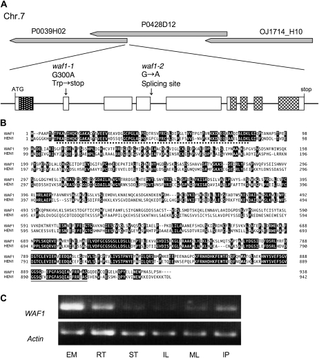
Molecular characterization of the WAF1 gene. A, Map position and structure of WAF1. Exons in pitted and checked boxes indicate the positions of the dsRNA-binding domain and the methyltransferase domain, respectively. Locations of the two mutations are indicated. B, Deduced amino acid alignments of WAF1 and HEN1. Dotted and solid lines indicate the dsRNA-binding motif and the methyltransferase motif, respectively. C, RT-PCR analysis of WAF1. EM, Embryos at 5 DAP; RT, roots; ST, stems; IL, immature leaves; ML, mature leaves; IP, young panicle at the primary rachis branch differentiation stage.

