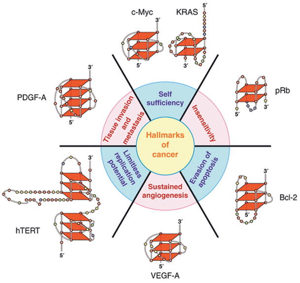
Fig. 1.

The six hallmarks of cancer [9] shown with the associated G-quadruplexes found in the promoter regions of these genes. As described in the text, the various G-quadruplexes differ by folding pattern, number of tetrads, loop size and constituent bases. In this a subsequent models, bases are colored as follows: guanine, red; cytosine, yellow; thymine, blue; adenine, green.
