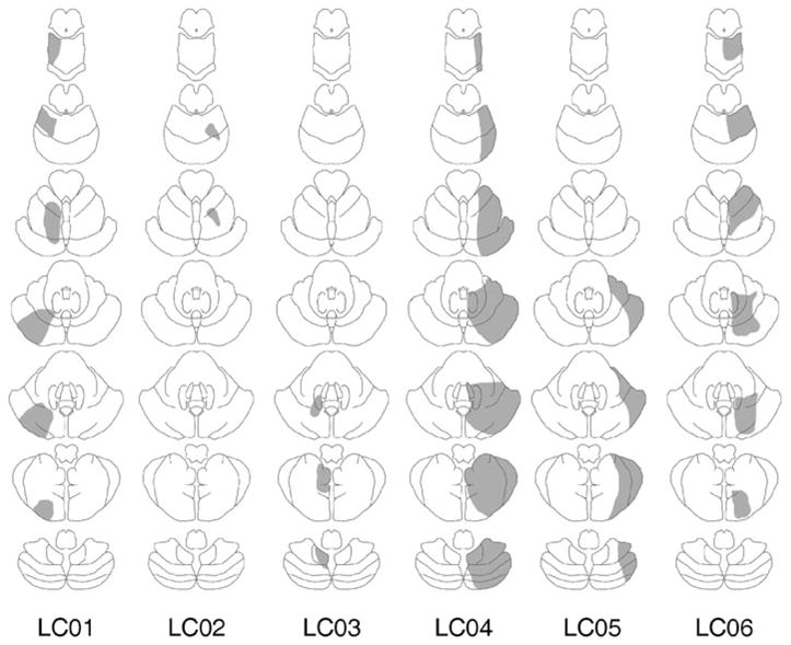
Fig. 2.

Lesion reconstructions (in gray) based upon computed tomography or magnetic resonance imaging for the patients with lateral cerebellar lesions. For each patient, the lesions are presented on a schematic of seven axial sections from superior (top) to inferior (bottom). LC, lateral cerebellar patients.
