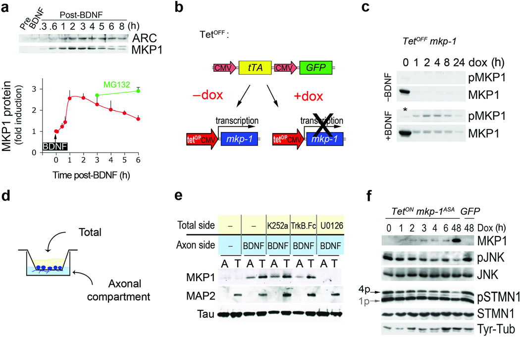
Figure 4. BDNF controls MKP-1 expression in a spatio-temporal manner.
(a) A 5-min pulse treatment with 50 ng/ml BDNF induces transient expression of MKP-1. Arc is shown for comparison. MKP-1 protein levels were expressed as fold induction compared to pre-stimulation (mean + s.d., n=3). MG132 was added just after the BDNF pulse for the indicated times. (b) Scheme of the tetOFF system composed of 2 plasmids used to uncouple MKP-1 expression from BDNF signaling. (c) MKP-1 protein turnover was rapid but prolonged by BDNF-induced phosphorylation in 293-TrkB cells when MKP-1 induction was uncoupled from BDNF signaling. Addition of doxycyclin (DOX) turned off MKP-1 expression. The * indicates that the sample was not treated with BDNF. (d) Schematic representation of the setup used to physically separate axons from the somatodendritic compartment. (e) Representative Western blots of the material scraped from both sides of the filter membrane. Pre-treatment of the total side with K252a, TrkB.Fc or U0126 exerted distinct effects on retrograde BDNF signaling and MKP-1 induction. Tau and MAP2 are markers of the axons and somatodendritic compartments, respectively. (f) To uncouple MKP-1ASA induction from BDNF signaling we used a tetON system where DOX induced MKP-1 in cortical neurons infected at DIV1–5. 1p and 4p indicate the mono- and multi-phosphorylated forms of STMN1, respectively.

