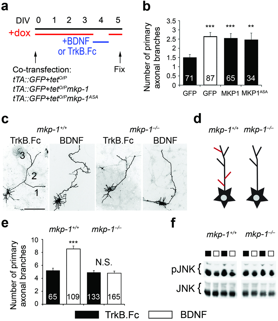
Figure 5. MKP-1 mediates BDNF-induced axon branching.
(a) Timeline of the experimental procedure used to manipulate axonal branching in vitro. (b) Number of primary axonal branches expressed as mean ± s.e.m. GFP/TrkB.Fc is significantly different from MKP-1 (***, p=0.0004, n=6 experiments), MKP-1ASA (**, p=0.0046, n=3 experiments) and GFP/BDNF (***, p<0.0001, n=5 experiments) using a t-test. (c,e) Images (c) and quantification (e) of the number of primary branches in GFP-transfected cortical neurons derived from mkp-1−/− mice and their wildtype littermates. Results from 3 mkp-1+/+ and 6 mkp-1−/− embryos were expressed as mean±s.e.m. The number of cells analyzed is indicated in the bars. BDNF induces branching in wildtype neurons only (wildtype, TrkB.Fc vs BDNF: p=0.0036; mkp-1−/−, N.S, not significant, TrkB.Fc vs BDNF: p=0.547 using a t-test). Scale bar, 50 µm. (d) Scheme summarizing the effects on branching. Cultured neurons grow axons and branch (black outline). This branching can be enhanced by BDNF or KCl (red lines) in wildtype neurons, however, mkp-1 null neurons fail to increase arborization in response to stimulation. (f) While incubation with BDNF for 3 h decreases pJNK levels in wildtype neurons (see also Fig. 3e,f), it fails to do so in mkp-1−/− cells.

