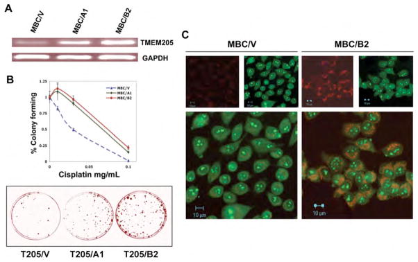
Fig. 5.
A: RT-PCR determination of expression levels of two individual clones isolated from TMEM205 (MBC)-transfected, and G418-selected stable clones. MBC/V was a control clone transfected with vector only. GAPDH served as a control. B, resistance levels of the TMEM205 transfectants, MBC/A1 and MBC/B2, to cisplatin; the upper panel shows the killing curves, and the lower panel shows methylene blue-stained colonies after exposure to cisplatin 0.1 ug/ml for 9 days. Each point was a mean of triplicate determinations on clonogenic assays after 9–10 days of exposure to the compound. Confocal images shown in C demonstrate reduced accumulation of F-CP (green) in TMEM205-transfectants, MBC/B2, double-stained with Rhodamine red-conjugated antibody directed to the TMEM205 polyclonal antibody (red).

