
Figure 9.
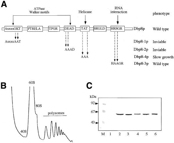
Site-directed mutagenesis of Dbp8p. (A) Four of the RNA helicase conserved motifs were individually altered by site-directed mutagenesis. The name of the mutant allele, the nature of the mutation and the associated growth phenotype are indicated. (B) Polysome analysis of MCD8-3C [pMCD8-11A] (dbp8-3) grown in YPD at 30°C. Twelve A260 units of cellular extract were resolved on a 7–50% sucrose gradient. Sedimentation is from left to right. The peaks of free 40S and 60S ribosomal subunits, 80S free couples/monosomes and polysomes are indicated. (C) Western blot analysis of the mutant proteins. Equal amounts of total protein extracts from MCD8H-3C [pMCD8-1] carrying, respectively, YCplac111 (lane 1), pMCD8-8 (ProtA–Dbp8p; lane 2), pMCD8-9B (ProtA–Dbp8-1; lane 3), pMCD9-10B (ProtA–Dbp8-2; lane 4), pMCD8-11B (ProtA–Dbp8-3; lane 5) and pMCD8-12A (ProtA–Dbp8-4; lane 6) were separated by 10% SDS–PAGE. The ProtA-tagged proteins were detected by western blot using rabbit immune serum and goat anti-rabbit alkaline phosphatase-conjugated antibodies. Pre-stained low range standard (Bio-Rad) was used as the standard for molecular mass determination.