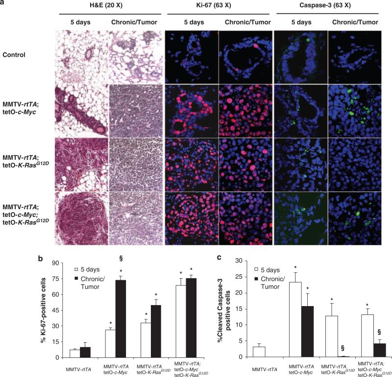
Figure 1.
Inducible expression of K-RasG12D and c-Myc in mouse mammary glands results in distinct histopathology, ectopic proliferation, and apoptosis. (a) Hematoxylin–eosin (H&E) staining of mammary gland cross-sections. The first two columns show that expressions of K-RasG12D or K-RasG12D and c-Myc result in dysplasia in mice treated with doxycycline for 5 days or until tumors developed (chronic means that controls were treated chronically for 10 months but did not develop tumors). The first column was from H&E performed in paraffin-embedded 10 mm sections; the second was performed in 10 μm frozen sections. All immunostainings (columns 3–6) were performed in 7–10 μm frozen mammary cross-sections. The second pair of columns show mammary epithelial cells immunostained with antibodies against Ki-67 in red (Abcam, Cambridge, MA, USA, ab15580; the secondary antibody is conjugated with Alexa Fluor 555). The third pair of columns show mammary epithelial cells immunostained with antibodies against cleaved-caspase-3 in green (Cell Signaling, Danvers, MA, USA, 9661; the secondary antibody is conjugated with Alexa Fluor 488). The nuclei were stained with DAPI in blue. (b, c) The percentages of proliferating cells (with positive Ki-67 staining) and apoptotic cells (with positive caspase-3 staining) were calculated at 5 days or in chronically induced mice. Each group included three independent mice. Averages and s.d. were calculated as 200 epithelial cells per mouse. Significance was assessed using an unequal variance t-test, calculated in Excel (Microsoft, Redmond, WA, USA) (significance: *P<0.05 as compared with MMTV-rtTA controls, §P<0.05 when comparing the same transgenic group 5 days with long-term treatment).

