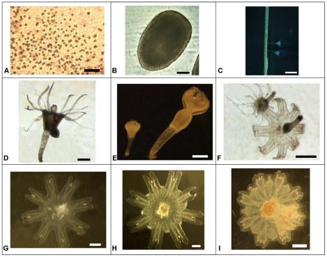
Figure 1. Cotylorhiza tuberculata. Various stages of its life history.
Pictures obtained during the simulation of Cotylorhiza tuberculata life cycle in the laboratory. A) Symbiotic zooxanthellae in the tissue of the mature medusae (scale 50 μm); B) Planula (scale 50 µm); C) Detail of scyphistomae on the leaves of the marine angiosperm Cymodocea nodosa (scale 2 mm); D) Budding scyphistoma (scale 500 μm); E) Scyphistoma initiating strobilation (scale 500 μm); F) Scyphistoma totally developed and scyphistoma about to liberate an ephyra (scale 500 μm); G) Ephyra (500 μm); H) Metaephyra (scale1 mm); I) Young medusa (scale 1 mm). Authors of the pictures: C. Lama: A; J.B.Ortiz: B; L.Prieto: C; D.Astorga: D–I.

