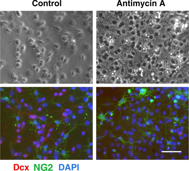
Figure 3.
Mitochondrial inhibition does not induce a significant change in the number of immature oligodendrocytes. Control (left panels) and antimycin A-treated (2 μm for 24 h) (right panels) cultures stained with NG2 (green) and Dcx (red) markers. Cell nuclei are counterstained with DAPI (blue). Antimycin A treatment promoted marked loss of Dcx+ cells but did not significantly change NG2 staining. Scale bar, 50 μm.

