
Fig. 2.
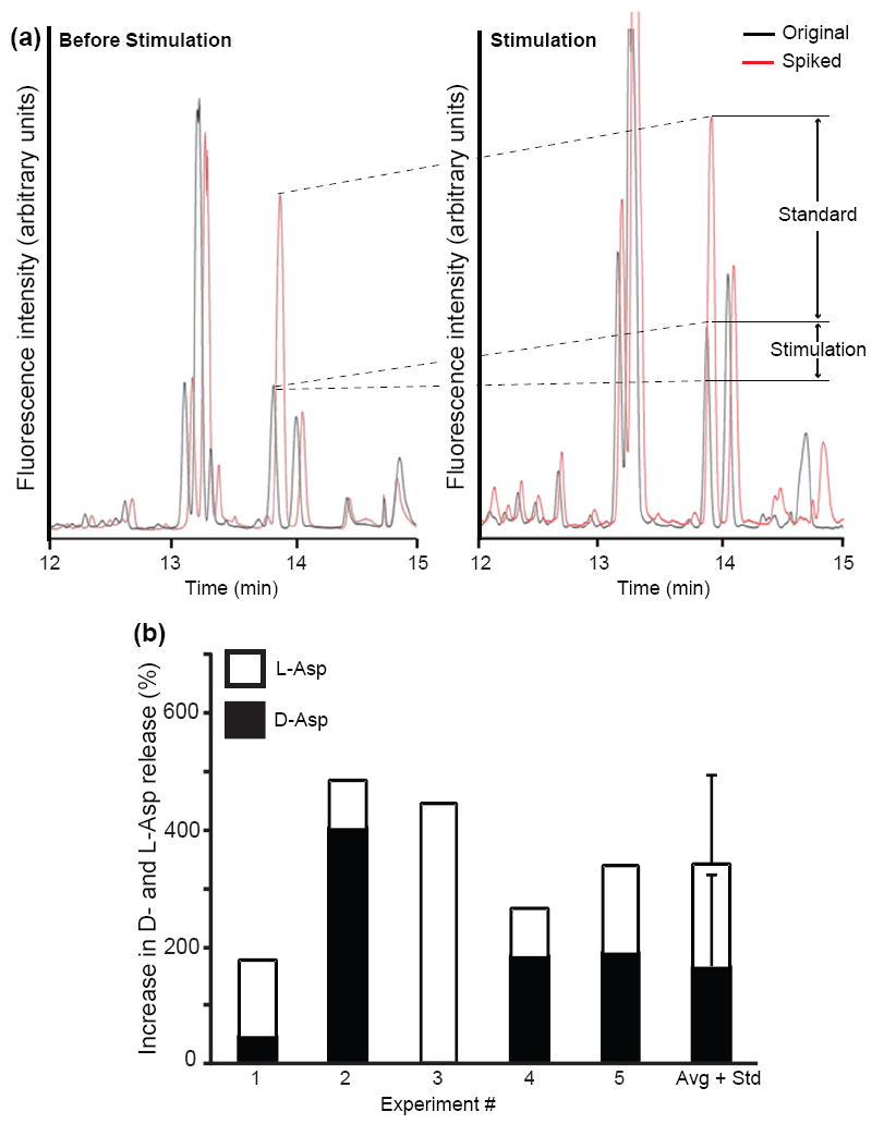
Elevated extracellular concentrations of potassium ions potentiate release of L- and D-Asp from the cerebral ganglia. CE-LIF electropherograms of cerebral ganglion releasate: (a) before KCl stimulation (left) and after KCl stimulation (right). In both cases, the black trace shows the original sample and the red trace shows the sample spiked with 1 μM D-Asp, used to calibrate the signal levels. Arrows indicate the increase in D-Asp signal due to the addition of standard and the change due to potassium ion stimulation (labeled standard and stimulation, respectively). (b) Stacked histograms summarizing results from five experiments, with statistical data placed in the sixth column. For each experiment, the extracellular potassium concentration was increased to 53 mM during the 20 min stimulation.