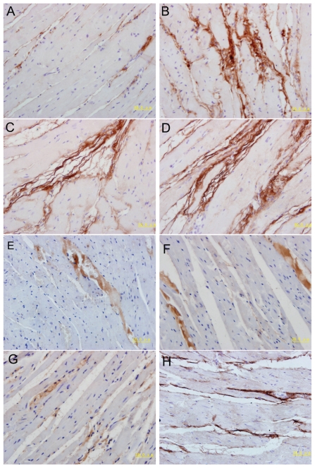
Figure 3.
Immunohistochemical staining of the myocardium with type I and III collagens in experimental rats. The top two panels show immunohistochemical staining of the myocardium with type I in the control (A), fructose (B), MS-GFP (C) and MS–IL-18 (D) rats (magnification 400×). The lower two panels show immunohistochemical staining of the myocardium with type III in the control (E), fructose (F), MS-GFP (G) and MS–IL-18 (H) rats (magnification 400×). An increase in type I and III collagen levels was observed in FFRs compared with control rats, and visible increases in type I and III collagen levels were noted in the MS–IL-18 rats. Bar = 50 μm.

