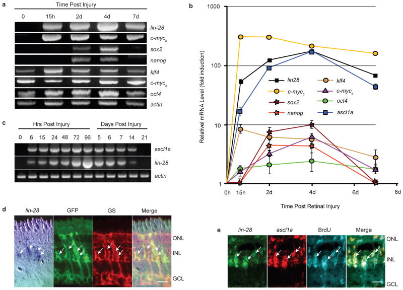
Figure 1.
ascl1a and lin-28 mRNAs are rapidly induced in dedifferentiating MG following retinal injury. (a) RT-PCR shows induction of pluripotency factor mRNAs following retinal injury. (b) Real-time PCR quantification of pluripotency factor mRNA levels during retina regeneration. Data represent means ± s.d. (n=3 individual fish; compared to uninjured retina, P=0.0001 or less for lin28, c-mycb, and ascl1a expression at all time points post retinal injury; P=0.0001 for klf4 at 15 hpi, and 2 and 4 dpi; P=0.02 for klf4 at 7 dpi; P=0.0001 or less for sox2, c-myca and nanog at 2 and 4 dpi; P= 0.0425 for c-myca at 7 dpi; P=0.0178 and 0.0069 for oct4 at 2 and 4 dpi, respectively). Note Y-axis is fold induction in log scale and normalized to 0 hr time point that is assigned a value of 1. (c) RT-PCR shows ascl1a and lin-28 are coordinately induced following retinal injury. (d) In situ hybridization and immunofluorescence shows lin-28 RNA co-localizes with 1016 tuba1a:gfp transgene expression in glutamine synthetase (GS)+ MG at 4 dpi. Scale bar is 10 microns. (e) lin-28 and ascl1a double fluorescence in situ hybridization and BrdU immunofluorescence show lin-28 and ascl1a are co-expressed in proliferating MG-derived progenitors at 4 dpi. Three hrs prior to sacrifice, adult fish, whose retina was injured 4 days earlier, received an intraperitoneal injection of BrdU. Scale bar is 10 microns. Abbreviations: ONL, outer nuclear layer; INL, inner nuclear layer; GCL, ganglion cell layer; BrdU, bromodeoxyuridine; GS, glutamine synthetase; GFP, green fluorescent protein.

