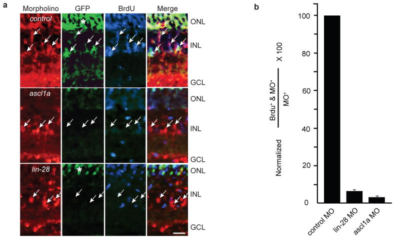
Figure 2.
Ascl1a and Lin-28 knockdown inhibit 1016 tuba1a:gfp transgene expression and MG-derived progenitor proliferation. (a) Control, ascl1a or lin-28 lissamine-tagged MOs were electroporated into the retina of 1016 tuba1a:gfp transgenic fish at the time of retinal injury and 3 hrs prior to sacrifice, at 4 dpi, fish received an intraperitoneal injection of BrdU. Arrows point to cells harboring lissamine-tagged MO. GFP and BrdU immunofluorescence show Ascl1a and Lin-28 knockdown suppress 1016 tuba1a:gfp expression and proliferation of MG-derived progenitors. (b) Quantification of the total number of MO+ cells that are in S phase as indicated by BrdU uptake. Data represent means ± s.d. (n=3 individual fish; compared to control MO, lin-28 MO and ascl1a MO P=0.000178 or less). (*) identifies autofluorescence in ONL. Scale bar is 10 microns and applies to all photomicrographs.

