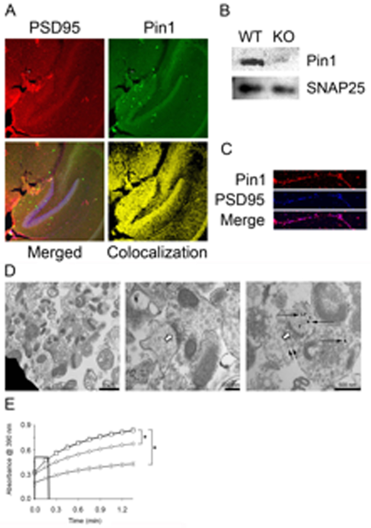
Fig. 1. Pin1 is present and active in post-synaptic terminals.

(A) Representative 100× confocal images of 30 µm hippocampal slices from P21 to P25 mice immunostained with anti-PSD95-Rhodamine, anti-Pin1-FITC, and To-Pro3. Merge panel shows uncolocalized (Pin1-red, PSD95-green, and To-Pro3-blue) and colocalized (yellow and magenta) points, whereas colocalization panel shows only points of Pin1 and PSD95 colocalization (yellow), (B) Pin1 and SNAP25 were detected by Western blot of 10 µg SN protein from WT and Pin1−/− mice. (C) Representative confocal image (600×) of E17 cortical neuron dendrites, 18 DIV. Cells were labeled with anti-Pin1-Rhodamine and anti-PSD95-Cy5 to identify colocalization (magenta). (D) EM of SN stained with anti-Pin1 coupled to gold beads [61]. Left and Center: vesicles (bars:1 µm left, 200 nm right) containing pre- and post-synaptic terminals. Right: representative region (bar 500 nm) stained with anti-Pin1-gold. Open arrows denote post-synaptic densities. Long, narrow arrows denote Pin1 staining. Small, narrow arrows denote synaptic vesicles. (E) SN were untreated (□), or pre-treated with 1 µM juglone (◊), or 1 µM CsA (○) for 10 min prior to lysis and isomerase activity assay. No SN control (X) contained complete reaction except SN, n=3, ± SEM, * denotes p<0.001. Initial slopes were calculated to determine K (see Table 1A).
