
Fig. 2. Pin1 suppresses dendritic translation.
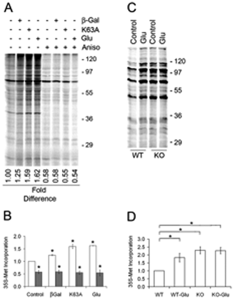
(A) SN were preincubated for 15 min. without (lanes 1–4) or with 40 µM anisomycin (Aniso, lanes 5–8) prior to addition of 35S-Met and no additional treatment, transduction with 50 nM TAT-βGal (βGal) or 50 nM TAT-Pin1-K63A (K63A), or treatment with Glu for 30 min prior to lysis and SDS-PAGE. (B) Total 35S-Met incorporation into protein was quantitated by phosphorimaging. Relative translation compared to control SN (□) was 1.25 ± 0.03 for βGal, 1.59 ± 0.09 for K63A and 1.62 ± 0.01 for Glu, 0.58 ± 0.06 for Aniso (■), 0.58 ± 0.07 for Aniso plus βGal, 0.55 ± 0.07 for Aniso plus K63A, 0.54 ± 0.12 for Aniso plus Glu, n=3, ± SEM, * denotes p<0.019 between K63A and βGal, p<0.003 between K63A and control. (C) WT or Pin1 KO SN were untreated (Control) or treated with Glu for 30 min. Total 35S-Met incorporation was quantitated (D) n=3, ± SEM, * denotes p<0.04.