
Fig. 3. Pin1 associates with signaling proteins, and Pin1−/− hippocampal slices show increased L-LTP.
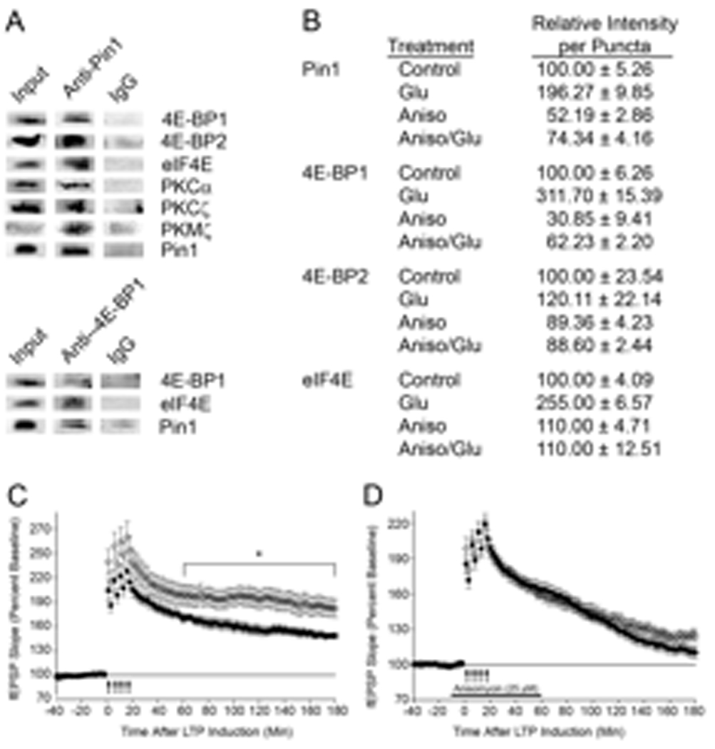
(A) WT brain homogenates (600 µg) were lysed and immunoprecipitated with pre-immune IgG, anti-Pin1, or anti-4E-BP1 followed by immunoblot, n=3. Homogenate (50–200 µg, input) was used as a positive control. (B) E17 cortical neurons, DIV18, were untreated (Control), stimulated with Glu, or pretreated with 40 µM Aniso before Glu, and then fixed, stained, and the dendrites visualized. Fluorescence of ~300 random puncta were quantitated and SEM determined. (C) L-LTP was induced in Pin1−/− (O; n=11 slices, n= 3 mice) or Pin1+/+ (●; n=9 slices, n=3 mice, p =0.0295) hippocampal slices with four trains of high frequency stimulation. (D) L-LTP was induced in anisomycin-treated hippocampal slices from Pin1+/+ [●; n = 12 slices, n = 3 mice, p = 0.0053 (WT with and without Aniso)] and Pin1−/− (O; n = 11 slices, n = 3 mice, p = 0.0003 (KO with and without Aniso)).