Fig. 1.

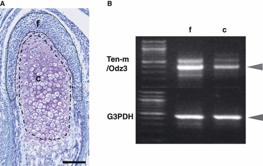
(A) Sagittal view of the mandibular condylar cartilage of a 1-day-old mouse stained with toluidine blue (TB). The two enclosed areas (f and c) show the division of the mandibular condylar cartilage. [This division was also used for laser-capture microdissection (LCM) and cDNA microarray analysis in our preliminary research.] f is the ‘fibrous layer’ not showing metachromasia, and c is the ‘cartilage layer’ showing metachromasia, as shown by TB staining. Bar = 100 μm. (B) Reverse transcription-polymerase chain reaction (RT-PCR) analysis of the expression of Ten-m/Odz3 in the two layers. RT-PCR was performed with total RNA samples from the fibrous (f) and cartilage (c) layers using LCM. The upper row shows Ten-m/Odz3 (396 bp), and the lower layer represents glyceraldehyde-3-phosphate dehydrogenase (G3PDH), a housekeeping gene. Ten-m/Odz3 expression was normalized to the expression of G3PDH. Results are representative of five independent experiments.
