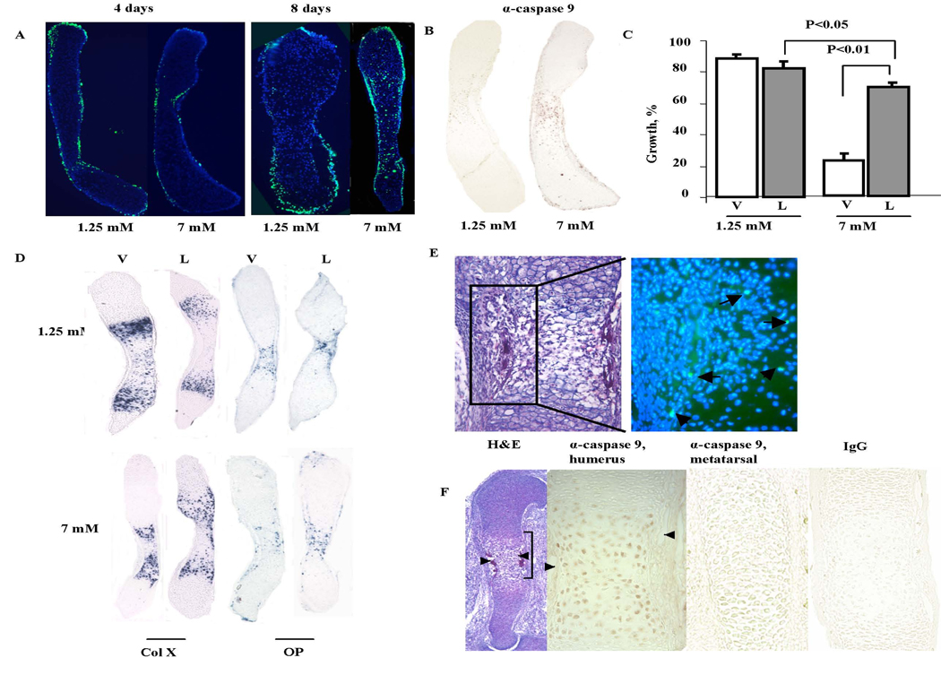
Figure 4.
Evaluation of the mitochondrial apoptotic pathway activation. A. TUNEL assays of metatarsals cultured with 1.25 mM and 7 mM phosphate were performed on sections of metatarsals cultured with 1.25 and 7 mM phosphate for 4 and 8 days. B. Immunohistochemistry with anti-cleaved caspase-9 antibody was performed on sections of metatarsals isolated after 4 days in culture with 1.25 mM and 7 mM phosphate. Non –specific IgG controls confirmed the specificity of the signal (data not shown). C. Percentage increase in the length of metatarsals cultured with 1.25 mM and 7 mM phosphate in the presence of LEHD-FMK (L) or vehicle (V) after 8 days in culture. D. In situ hybridization analyses were performed on sections of metatarsals cultured with 1.25 and 7 mM phosphate for 8 days with 20 µM LEHD-FMK (L) or vehicle (DMSO) (V). E. H&E staining and TUNEL assay of e15.5 humerus. The fluorescent image of the boxed area on the left is shown in the merged DAPI (nuclear stain) and FITC (TUNEL stain) image on the right. Arrows point to TUNEL positive hypertrophic chondrocyte nuclei. F. Cleaved caspase-9 immunoreactivity is observed in vivo in hypertrophic, but not proliferative chondrocytes, in the humerus of e14.5 mice. No immunoreactivity is observed in the hypertrophic chondrocytes of metatarsals of e14.5 mice. Arrows point to blood vessels. Data are representative of that obtained from at least 3 independent experiments.

